An echocardiogram is a cutting-edge diagnostic test that uses ultrasound waves to create images of your heart. It provides valuable information about the structure and function of the heart, including the size of the chambers, the thickness of the heart walls, and the movement of your heart valves. An echocardiogram can also assess the blood flow through the heart and identify abnormalities or blockages. You may benefit from an echocardiogram Upper East Side if you experience unexplained chest pain or shortness of breath.
Transthoracic echocardiogram
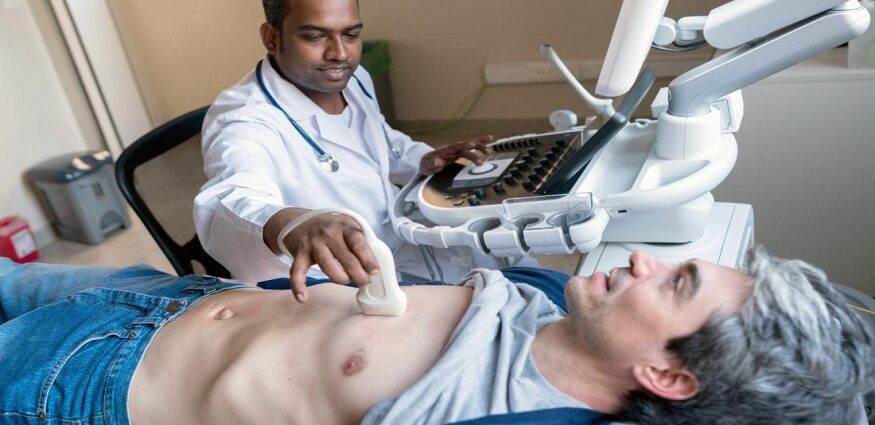
Transthoracic echocardiogram (TTE) is a non-invasive imaging technique that utilizes high-frequency sound waves to generate images of your heart. During a TTE, your provider places a small transducer on your chest wall, which emits sound waves that bounce off your heart muscle and create detailed images of its structures and functions. This type of echocardiogram can provide information on the size and appearance of your heart, the movement and thickness of its walls, and the functioning of its valves and blood vessels.
Transesophageal echocardiogram
Transesophageal echocardiogram (TEE) uses a specialized probe, which your doctor inserts into your esophagus, to obtain detailed images of your heart. The esophagus is located behind the heart, which allows for clearer and more accurate images than a standard echocardiogram. Medical experts often use TEE to evaluate the structure and function of the heart, diagnose heart conditions such as atrial fibrillation or infective endocarditis, and guide interventions such as atrial septal defect closure. They conduct the procedure under sedation. TTE is generally safe, although there are some risks, such as throat irritation or bleeding.
Doppler echocardiogram
A Doppler echocardiogram produces detailed images of your heart and blood vessels using ultrasound waves. The Doppler component of this test allows your doctor to evaluate blood flow in your heart and blood vessels by measuring the frequency and velocity of the sound waves as they bounce off of moving blood cells. They can use this information to diagnose various heart conditions, including valve disorders, and to monitor the effectiveness of treatments.
Stress echocardiogram
A stress echocardiogram evaluates the function of your heart during physical stress. During this procedure, the patient exercises on a treadmill or stationary bike while their heart rate and blood pressure are monitored. Once you reach your target heart rate, your provider uses a specialized ultrasound machine to capture images of your heart at rest and during stress. They then analyze the images to assess your heart’s response to stress and detect any abnormalities in its function.
When do you need an echocardiogram
An echocardiogram is a non-invasive test that utilizes ultrasound waves to provide detailed images of the heart’s structure and function. It can diagnose and monitor heart conditions such as heart disease, heart failure, valve problems, and congenital heart defects. Your physician may recommend an echocardiogram if you experience chest pain, shortness of breath, palpitations, swelling in your legs, or if you have risk factors for heart conditions, like high blood pressure or high cholesterol. Additionally, an echocardiogram can assess treatment effectiveness or guide certain heart procedures. Ultimately, the decision to perform an echocardiogram will depend on your medical history, symptoms, and other diagnostic test results.
Call the Upper East Side Cardiology office or book an appointment online for more echocardiogram information.