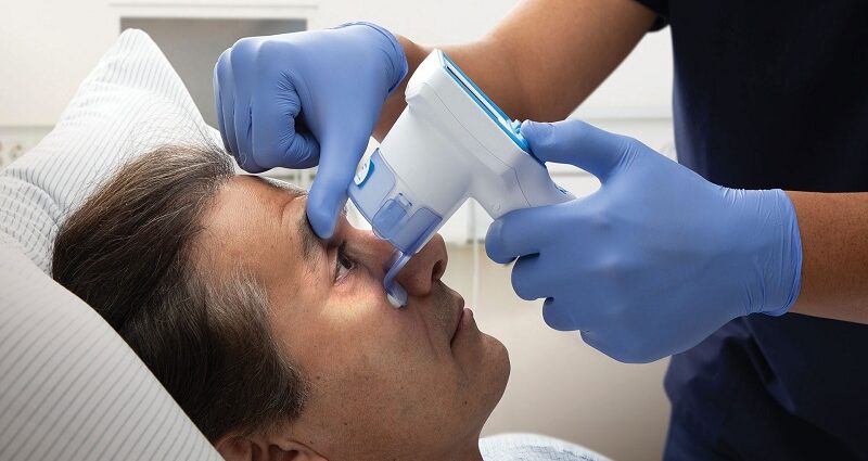
Pupil diameter measurement is an important component of neurological assessment; it gives a view into the neurological status of the patient. Modern technology has provided new methods that help in increasing the accuracy of these measurements. This blog discusses such advanced techniques and underlines their relevance in the field of neurology.
The Importance of Pupil Diameter Measurement
Diameter Measurement
Pupil diameter measurement is one of the most important assessments of neuro exams that provides important data about the ANS and brain activity. Abnormalities in pupil size may signify a wide range of neurological disorders, including, traumatic brain injuries, and neurodegenerative diseases among others. It is for this reason that accurate and efficient measurement tools are crucial to neurologists and other healthcare givers.
Classic Approaches of Measuring Pupil Diameter
In the past, the assessment of pupil diameter was done with simple approaches such as the penlight test and simple rulers or pupil gauges. Although these methods are rather simple, they are not very accurate and do not allow us to carry out highly differentiated neurological examinations. They are also associated with inter-observer variability and thus increase the variability in the kind of care that a patient receives.
Progress in Pupil Diameter Measurement
Automated Pupillometry
Automated pupillometry is a revolutionary progress in the field of pupil diameter assessment. This technique uses the infrared system to capture the changes in the pupils and their dimensions. Equipment like the Neuroptics Pupillometer (NPi) gives out numeric results, which reduces the interference of the observer and boosts the diagnostic precision.
Percentage Change in Pupil Diameter
Among the parameters assessed with the help of automated pupillometers, one of the most vital is the percentage of the change in the pupil’s diameter. This parameter relates to the light stimuli and measures the attitude of the pupil, which gives information about the functioning of the autonomic nervous system. Assessment of the percent change in pupil size is very essential when diagnosing Horner’s syndrome, Adie’s pupil, and in the assessment of the severity of some conditions.
Integration with Neurological Tools
Automated pupillometry as a technique is valuable because the tool augments the range of neurological tests. For example, together with the results of pupillometry, it is possible to use EEG and MRI for a more detailed examination of a patient’s neurological condition. This multiple approach to diagnosis helps in increasing the chances of accurate diagnosis and better treatment plans.
Constriction Velocity
Another parameter is constriction velocity which refers to how fast the pupil constricts in response to light. This metric is especially valuable for the evaluation of the optic nerve and oculomotor pathways’ health. It is effective in neurological disorders’ early identification due to its ability to deliver accurate constriction velocity readings.
The Role of NPi in Neurological Assessments
The Neurological Pupil Index (NPi) is the total score of several parameters such as pupil size, pupillary response, and changes in size. Due to the high variability of pupillary size, the NPi can be considered a standardized index of pupillary function that interprets the pupillometry results more manageably. NPi score above 135 is usually normal for pupils while low scores may be an indication of neurological problems.
Applications of NPi
The NPi has multiple clinical uses: In the intensive care practice it is applied to assess ICU patients with head injuries and to inform management procedures and prognosis. In the case of outpatient neurology, the NPi plays a vital role in the diagnosis and treatment of diseases such as multiple sclerosis, Alzheimer’s disease, and Parkinson’s disease among others. The latter proves its versatility within various clinical settings that demand the integration of progressive pupillometry into basic neurological evaluations.
Future Directions
The prospect of pupil diameter measurement in neurology is quite bright and further research is being carried out to fine-tune these procedures. The portable pupillometers and merging of neuro exams with AI algorithms have the potential to transform neuro exams. Automated analysis of the pupillometry data could mean having real-time diagnostic support which would certainly help clinicians to make faster and more accurate decisions.
Conclusion
New techniques of measuring pupil diameter are a rather revolutionary step in the field of neurology as they provide more accurate and efficient methods of evaluating the state of neurological functions. Automation of pupil examination, the change in the size of the pupils in percentage, the speed of their constriction, and the NPI are an essential part of today’s neurological examinations. These techniques will become even more significant in the future with modern technologies, helping to diagnose and treat neurological disorders in patients.

