
As its name suggests, the temporomandibular joint (TMJ) is found at the junction of the temporal bone of the skull and the lower jaw (mandible), on either side of the face, in front of the ears. Each human therefore has 2 ATMs.
ATMs play a main role in essential functions involving the opening and closing of the mouth, namely speaking , chewing , swallowing and even yawning . They are therefore the most stressed joints in the human body.
In addition, they can perform front-to-back (anteroposterior) glides and pivots, in addition to slight left-to-right movements, in a synchronized fashion. All these features give them the title of the most complex joints in the human body.
Discover orthodontics as a treatment for temporomandibular joint (TMJ) disorders
THE COMPONENTS OF ATM
ATM is made up of several elements, the main ones being:
the base of the skull where the temporal bone is located and the condyle of the mandible , which is the small ball at the end of the ascending branch of the lower jaw bone;
small muscles that allow the mandible to move;
several ligaments and tendons ;
blood vessels and nerves;
articular surfaces between which an articular disc , also called a meniscus, is positioned. This disc serves as a small cushion between the condyle and the skull during the movements of the mandible in order to avoid any friction of the articular surfaces. It also helps to absorb and redistribute strong masticatory forces. This meniscus also has the particularity of following the mandible in the articular fossa during the movement of the jaw;
synovial fluid to ensure smooth jaw movement.
The auditory canal lies posterior to the ATM.
These elements, along with other surrounding structures and those attached to them, must be in perfect balance to function well.
THE MECHANICS BEHIND ATM MOVEMENTS
During the opening and closing of the mouth, the structures of the ATM are all solicited.
NORMAL OPENING AND CLOSING OF THE MOUTH
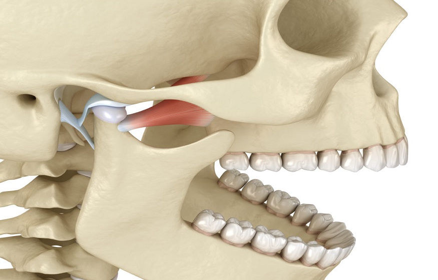
When the lower jaw is at rest, the condyle of the mandible (in beige) normally rests in the articular fossa and the articular disc (in pink), also called meniscus, is located between the condyle and the bone of the skull.
Note that the black circle behind the temporomandibular joint is the ear canal.
When the mouth is open, the condyle (in beige) lowers, moves forward and exits the articular fossa. Normally, the meniscus (in pink) also moves forward at the same time with the help of the ligaments (in red) which stabilize it and the TMJ muscles (small black lines in front of the condyle) to ensure comfort of movement without making noise or causing pain.
When the jaw joint is functioning normally, no noise should be heard when the person opens or closes their mouth. This also requires the synchronicity of the movements of the 2 ATMs.
However, sometimes the TMJ structures do not work in harmony and abnormal displacement of the articular disc may occur and noises may be heard, with or without pain.